Adrenergic receptors
How catechol amines act?
Catechol amines act as important chemical mediators in sympathetic system, but how they act?
Just like with acetylcholine, these catechol amines also produces pharmacological response by interacting with corresponding receptors. In the previous article, we have discussed that sympathetic system produce vasoconstriction at systemic blood vessels and vasodilatation at skeletal blood vessels.
How two quite opposite actions are possible with same mediator?
Again the answer is similar with that one we have discussed in cholinergic system. These mediators don't act on single receptor but act on two different receptors each producing different set of actions.
For example, epinephrine acts on α-receptors to produce vasoconstriction and β receptors to produce vasodilatation.
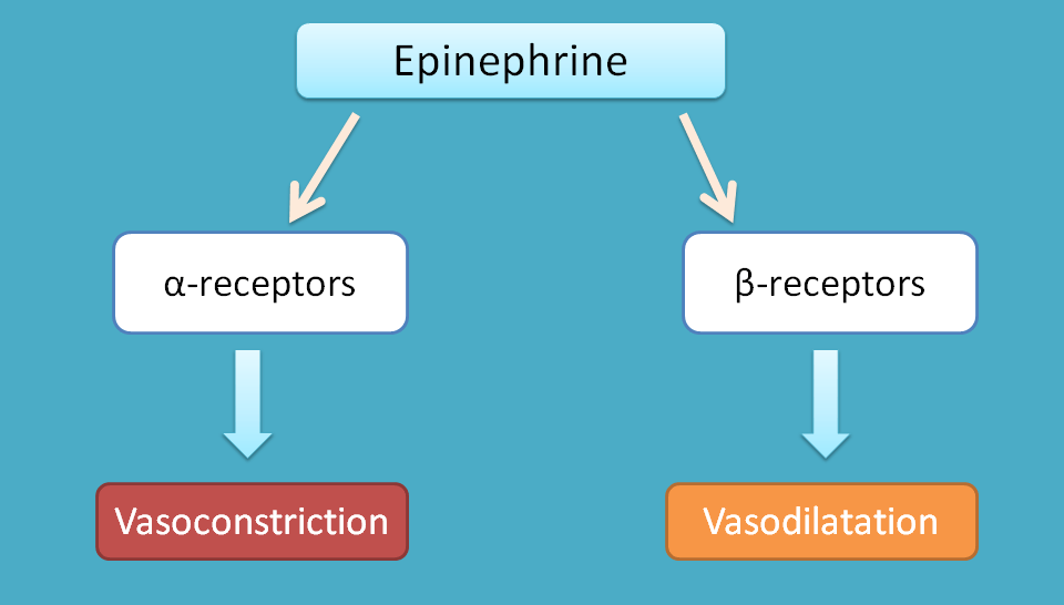
This can be further confirmed by dale’s vasomotor reversal.
Dale’s vasomotor reversal
It is a simple demonstration of existence of adrenergic receptors on vascular smooth muscle. Two types of adrenergic receptors such as α1 and β2 are present on vascular smooth muscle. α1 is responsible for vasoconstriction while β2 for vasodilatation. Under normal conditions, catechol amines preferentially bind to α1 receptors producing vasoconstriction.
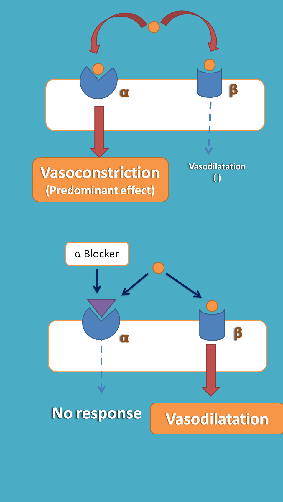
So, if α1 receptors are blocked by an alpha blocker, catechol amines act on β2 receptors producing vasodilatation. This reversal of response i.e. vasoconstriction to vasodilatation produced by alpha blocker is called as Dale’s vasomotor reversal.
This experiment again confirms that catechol amines don’t act on single receptor instead act on two different receptors. Based on this adrenergic receptors are classified into two categories
- Alpha receptors
- Beta receptors
Alpha adrenergic receptors can be classified into two types
- α1receptors
- α2receptors
Similarly beta receptors are classified into β1, β2 and β3 receptors.
So, let’s start our discussion with alpha adrenergic receptors in the next section.
35a. α–Adrenergic receptors
We have already discussed that alpha receptors are further sub classified into α1 and α2. Here we will discuss the location and function of these receptors.
α1–Adrenergic receptors
Location
Three important locations of α1–adrenergic receptors are
- Smooth muscle
- Glands
- Salivary gland
- Liver
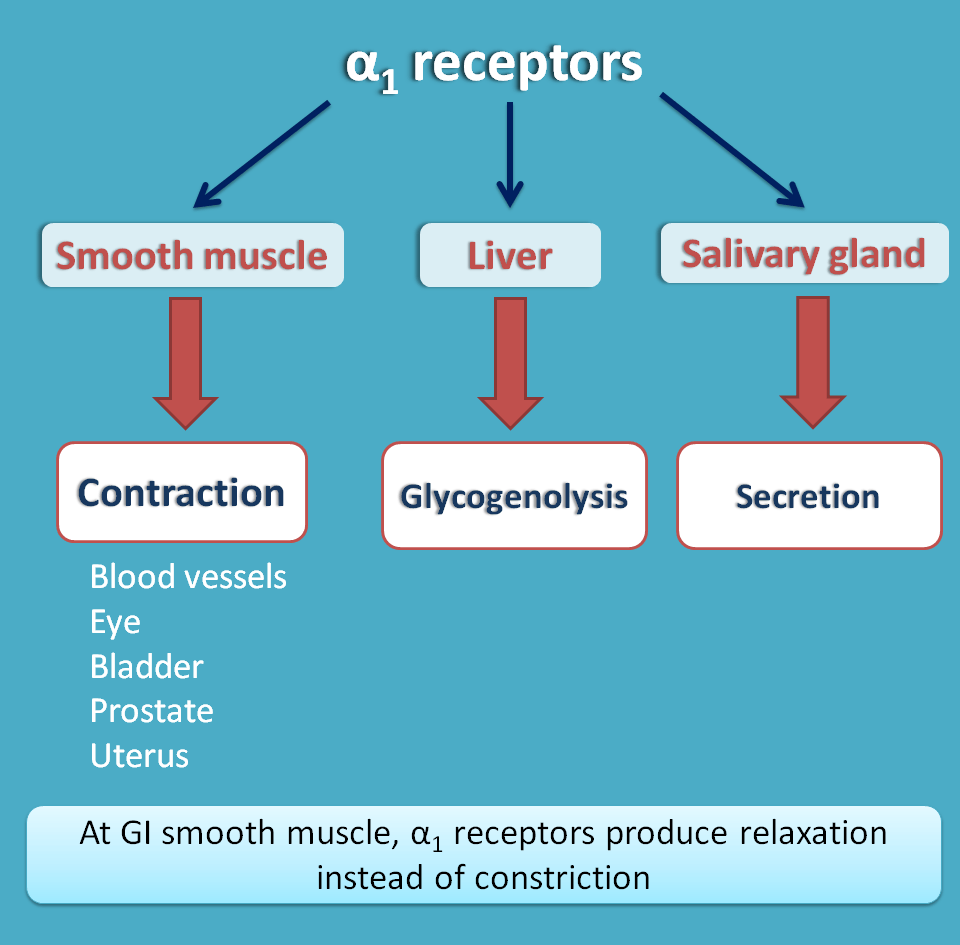
These receptors are present at various smooth muscles like
- Vascular smooth muscle
- Eye
- Bladder
- Prostate
- Uterus
- GI smooth muscle
Apart from smooth muscle, these receptors are also present on salivary glands and liver responsible for secretion and glycogenolysis respectively.
Function
They are G-protein coupled receptors of Gq type coupled with activation of phospholipase C. This activated phospholipase C then cleaves phosphatidylinositol biphosphate into two secondary messengers
- Inositol triphosphate (IP3)
- Diacyl glycerol (DAG)
IP3 is responsible for release of calcium from sarcoplasmic reticulum by acting on IP3 receptors.
DAG is responsible for increased calcium entry into the smooth muscle.
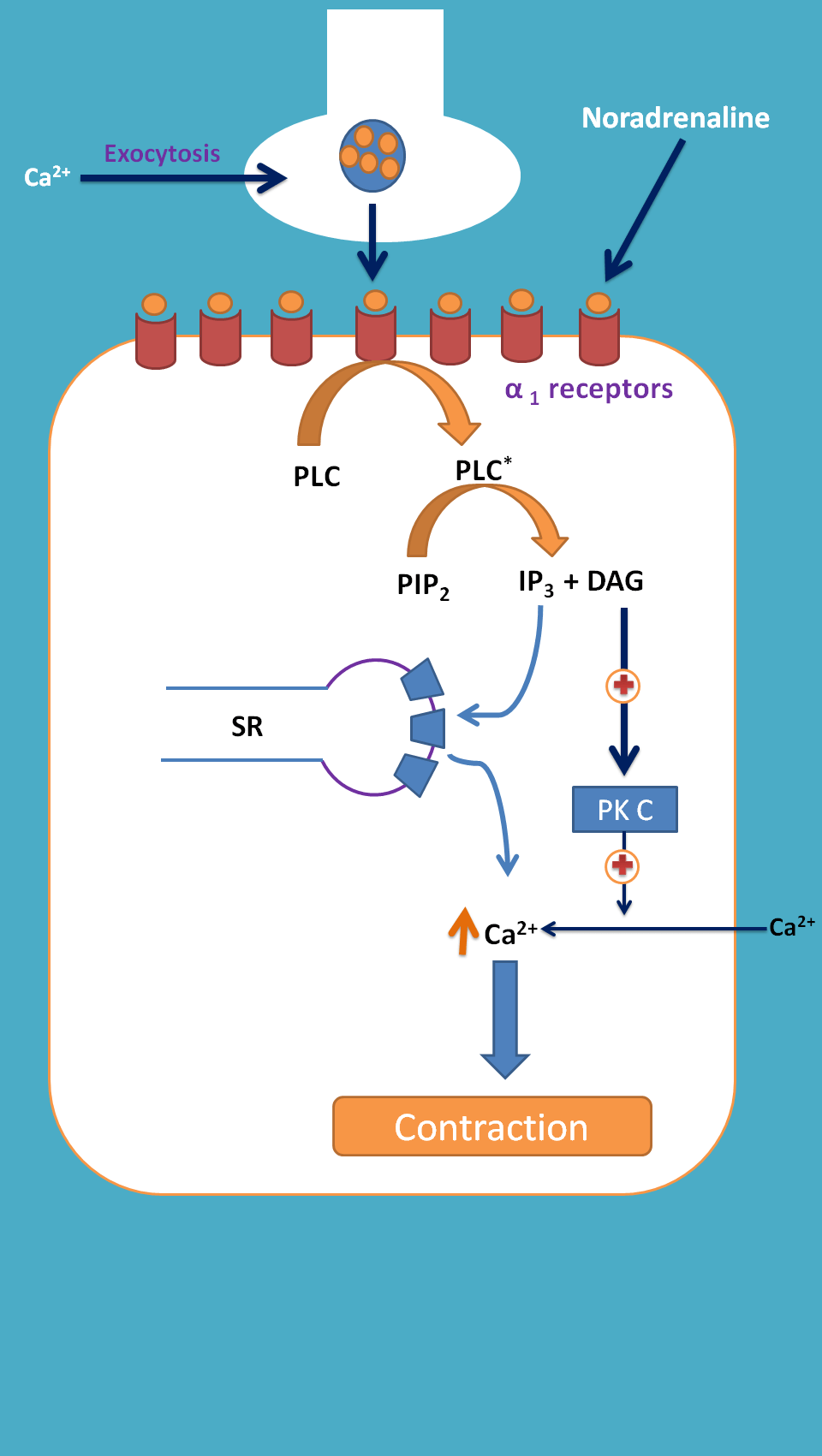
In this way, both IP3 and DAG are responsible for increased intracellular calcium leading to contraction of the smooth muscle.
Exception at GI smooth muscle
The α1 receptors at GI smooth muscle are again G-protein coupled receptors but not coupled with IP3/DAG instead coupled with opening of potassium channels leading to hyperpolarisation. Hence α1 receptors produce relaxation of GI smooth muscle decreasing motility.
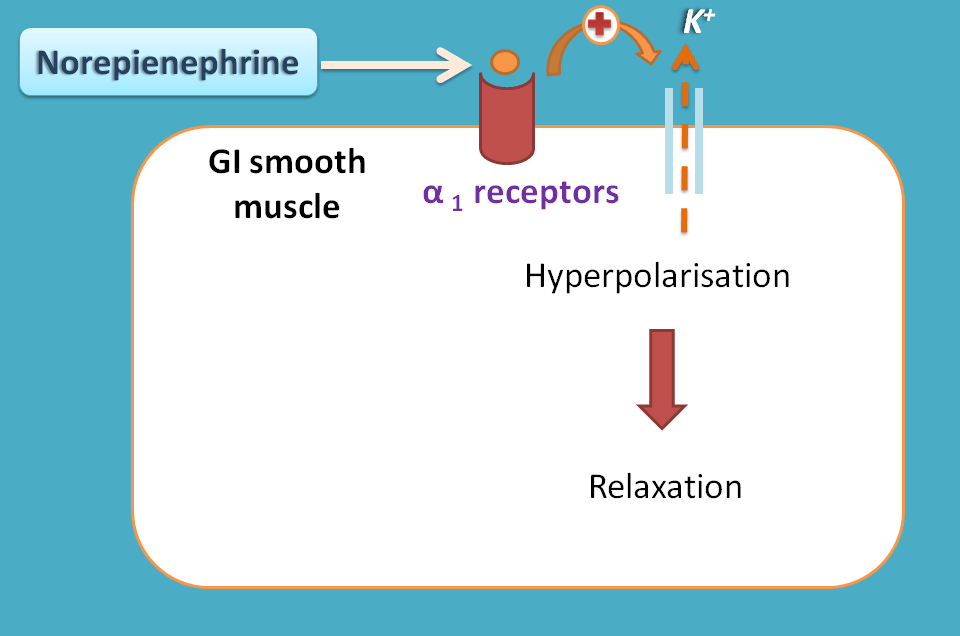
In summary we can conclude α1 actions as
- All smooth muscles are contracted except GI smooth muscle
- Secretion of salvia
- Glycogenolysis in liver
α2 adrenergic receptors
Location
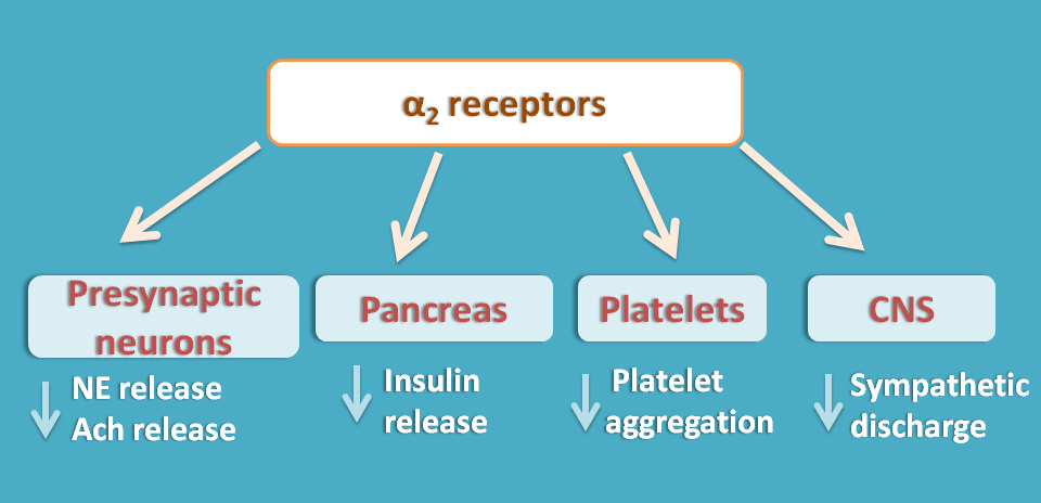
α2 receptors are mainly present as presynaptic at
- adrenergic neurons
- cholinergic neurons
- Beta cells of pancreas
They also present at
- Platelets
- CNS
Function
α2 adrenergic receptors are again G-protein coupled receptors coupled with inactivation of adenylyl cyclase. This results in decreased release of cAMP hence intracellular calcium. As calcium levels fall, the release of neurotransmitter from nerve terminal by exocytosis is inhibited.
At adrenergic neurons they play important role as auto inhibitory feedback mechanisms controlling norepinephrine release. At platelets they inhibit platelet aggregation and within the CNS they decrease central sympathetic discharge.